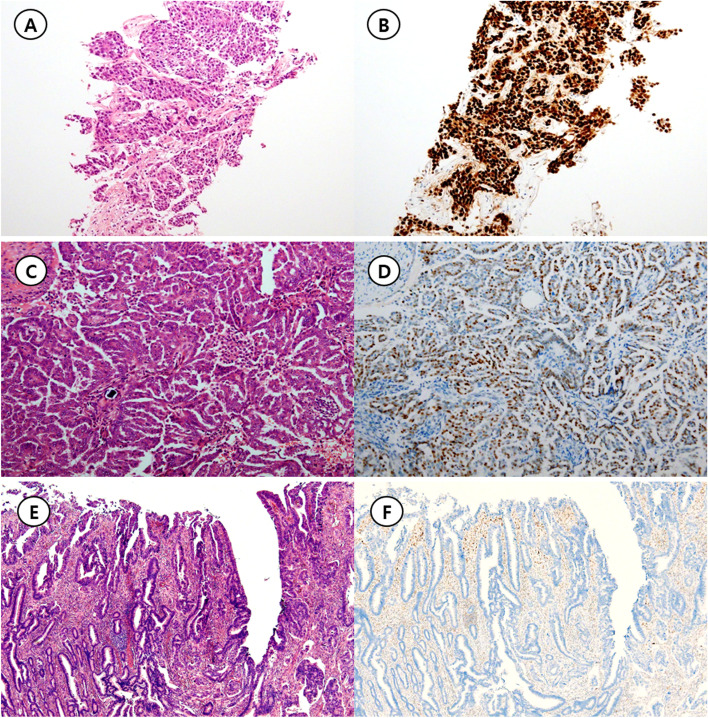
Fig. 1.
Representative figures for three p53 immunohistochemistry patterns. A Invasive ductal carcinoma of the breast harboring TP53 H193R mutation (Hematoxylin & Eosin, × 1.25 objective lens). B Diffuse strong immunoreactivity for p53, that is overexpression pattern (p53 immunohistochemistry, × 1.25 objective lens). C Low grade papillary serous carcinoma of the ovary without any oncogenic TP53 mutation (Hematoxylin & Eosin, × 10 objective lens) D Approximately a half of the tumor cells express p53 in the nuclei, that is usual pattern (p53 immunohistochemistry, × 10 objective lens). E Adenocarcinoma of the colon harboring TP53 H214Qfs*2 mutation (Hematoxylin & Eosin, × 4 objective lens). F Tumor cells are completely negative for p53 protein expression. Adjacent non-neoplastic cells showing p53 expression serves as an internal positive control. This pattern is classified as null pattern (p53 immunohistochemistry, × 4 objective lens)

