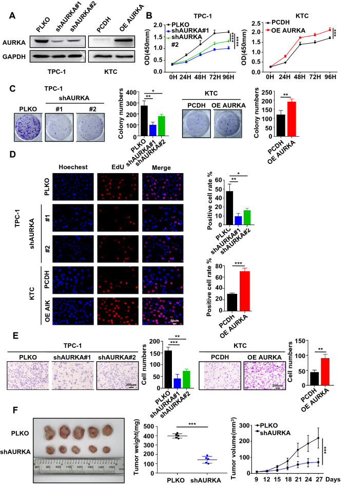
Fig. 2.
AURKA acts as an oncoprotein in PTC. A The protein level of AURKA was detected in AURKA knockdown TPC-1 cells and AURKA-overexpressing KTC cells by western blotting. B The viability of shAURKA-transfected TPC-1 cells or AURKA-overexpressing plasmid-transfected KTC cells compared with control cells was detected using the CCK-8 assay. C The shAURKA-transfected TPC-1 cells and AURKA-overexpression plasmid-transfected KTC cells were cultured for 10 days prior to crystal violet staining. D The ratio of EdU-labelled cells to total cells was observed in AURKA knockdown TPC-1 cells and AURKA-overexpressing KTC cells compared with the control group (original magnification × 20). E Transwell migration assays were used to detect the migration ability of AURKA knockdown TPC-1 cells and AURKA-overexpressing KTC cells compared with control cells. F Representative images of subcutaneous xenografts in nude mice derived from PLKO-transfected TPC-1 cells and shAURKA-transfected TPC-1 cells. n = 5 mice per group. Data are shown as the mean ± SD of three replicates (*P < 0.05, **P < 0.01, ***P < 0.001)

