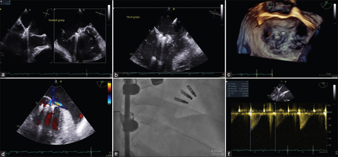
Figure 3.
Transthoracic echocardiogram image at grasping view showing placement of second MitraClip XTR lateral to first partially detached clip. (a) And the third Mitraclip XTR medial to the first partially detached clip. (b) 3D transthoracic echocardiogram image without. (c) And with (d) color Doppler and RAO Caudal fluoroscopic view (e) showing the final results with stable MitraClip devices. Continuous wave Doppler showing a mean gradient of 4 mmHg at the end of the procedure (f). RAO: Right anterior oblique

