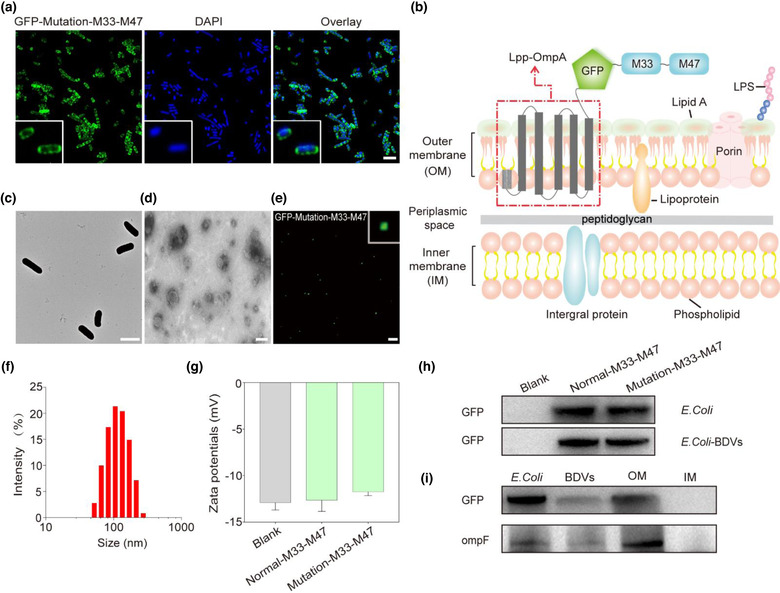
FIGURE 1.

Characterizations of GFP‐Mutation‐M33‐M47 E. coli BDVs. (a) Establishment of E. coli stably expressing GFP (Scale bar: 2 μm). (b) Schematic diagram illustration the presentation of neoantigens using Lpp‐OmpA system on E. coli outer membrane. (c) The TEM images shown the size and morphology of GFP‐Mutation‐M33‐M47 E. coli (Scale bar: 2 μm). (d) The TEM images shown the size and morphology of GFP‐Mutation‐M33‐M47 E. coli BDVs (Scale bar: 100 nm). (e) The confocal image of GFP‐Mutation‐M33‐M47 E. coli BDVs (Scale bar: 5 μm). (f) The size distribution of GFP‐Mutation‐M33‐M47 E. coli BDVs measured by dynamic light scattering (DLS). (g) The zeta potential of the E. coli BDVs. Blank (BL21(DE3)plysS): non‐heat shock transformation control, worked as a negative control. (h) Western blot analysis indicated the expression of GFP on E. coli and BDVs. (i) Western blot analysis indicated the expression of GFP and ompF on E. coli, BDVs, OM and IM
