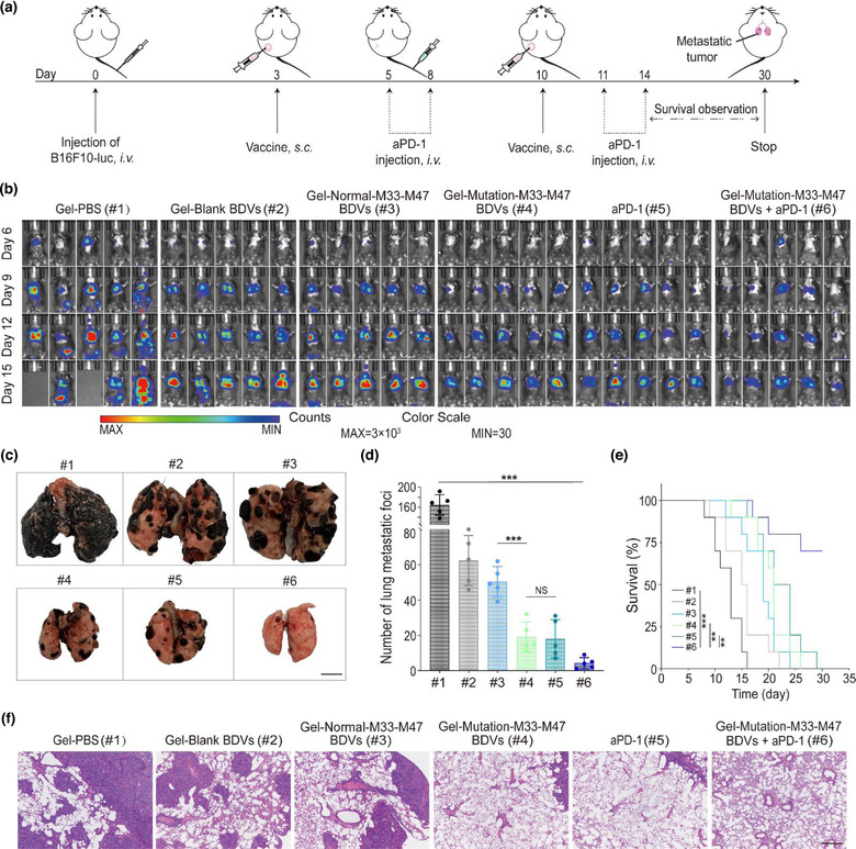
FIGURE 5.

In vivo suppression of antimetastatic tumour effect of BDV‐neoantigen vaccine. (a) Schematic illustration of the therapy of BDV‐neoantigen vaccine in B16F10‐luc model of metastasis. (b) In vivo bioluminescence images of the B16F10‐luc lung metastasis in different groups (n = 5). (c) Representative images of the lung metastatic nodules (Scale bar: 5 mm). (d) Numbers of lung metastatic nodules in different groups (n = 5), Error bar, mean ± s.d.. (e) Survival curves of different groups (n = 10). Day 0 is the day of tumour cells injection via tail vein. (f) H&E‐stained lung slices (Scale bar: 100 μm). (#1) Gel‐PBS, (#2) Gel‐Blank BDVs, (#3) Gel‐Normal‐M33‐M47 BDVs, (#4) Gel‐Mutation‐M33‐M47 BDVs, (#5) aPD‐1, (#6) Gel‐Mutation‐M33‐M47 BDVs + aPD‐1. NS: no significant, *P < 0.05, **P < 0.01, ***P < 0.001. One‐way ANOVA with Tukey post‐hoc tests (d) or the Long‐Rank (Mantel‐Cox) test (e)
