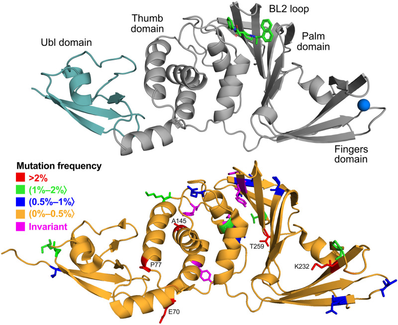
Fig 1. Structure of PLpro and location of the mutations.
Upper panel: cartoon representation of the protein with inhibitor S43 shown in sticks and zinc ion as a sphere (based on PDB ID: 7e35). Teal region of the protein indicates N-terminal ubiquitin-like domain (Ubl). Lower panel: cartoon representation of the protein with residues colored according to their mutation frequency (percentage of sequences with a given mutation out of all mutated sequences).

