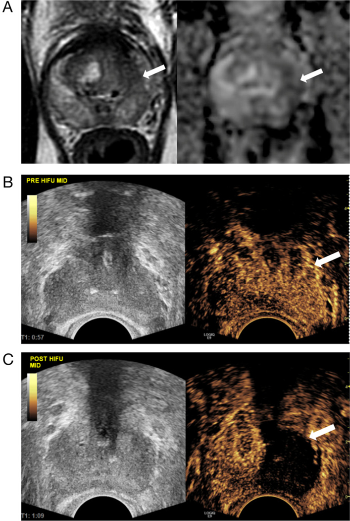
Figure 1.
Example of prostate MRI before HIFU and contrastenhanced TRUS appearance of prostate hemiablation with HIFU. A, T2-weighted (left) and ADC (right) MR images show a 1.2-cm left peripheral zone lesion in the 1- to 3-o’clock position, with low T2 and ADC signals (arrows), Prostate Imaging Reporting and Data System category 4. This appearance corresponded to Gleason 3 + 4 prostate cancer on biopsy. B, Split-screen grayscale (left) and contrast-enhanced TRUS (right) images before HIFU ablation. The left prostate lobe shows diffuse slightly increased enhancement (arrow) relative to the right. C, Split-screen grayscale (left) and contrast-enhanced TRUS (right) images immediately after HIFU ablation. The left prostate lobe shows complete absence of enhancement (arrow). The right prostate lobe enhances normally.

