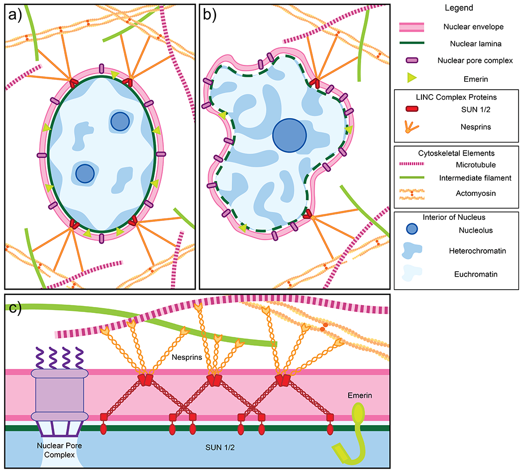
Figure 1.

a) Schematic of a normal nucleus. The nucleus is enclosed by the nuclear envelope (pink), which consists of an inner and outer lipid membrane. The inner membrane is studded with numerous proteins including LEM domain proteins such as emerin (light green). Other protein complexes span both the inner and outer membranes, including nuclear pore complexes (purple), which facilitate nucleocytoplasmic transport, and the LINC complex, comprised of SUN1/2 (red) and nesprins/KASH proteins (orange), which transmits mechanical force from the cytoskeleton to the nucleus. The lamina (dark green), a fibrillar mesh network, provides structure and support to the nucleus. Peripheral heterochromatin (dark blue regions) that is compacted and less accessible for transcription is anchored to the nuclear lamina. Euchromatin (light blue regions) are less compacted and more accessible for transcription. Normal cells usually have two to three nucleoli (dark blue circles), providing sites for ribosome assembly. b) Schematic of a cancerous nucleus and examples of changes to nuclear structure and organization, as observed in human samples, which may contribute to abnormalities in size and shape. Cancerous nuclei in many tissue types exhibit downregulation of a number of proteins including emerin, SUN1/2, and nesprins. In contrast, nucleoporins may be upregulated. Different cancer types are characterized by abnormal expression of nuclear lamins. Finally, chromatin organization is also often disrupted in nuclei of cancerous cells. c) A schematic of a cross-section of the nuclear envelope. Shown in greater detail is the structure of the LINC complex. Two SUN trimers and three KASH dimers interact via SUN-KASH 6:6 complexes which serve as nodes for force transduction and distribution (Gurusaran and Davies 2021).
