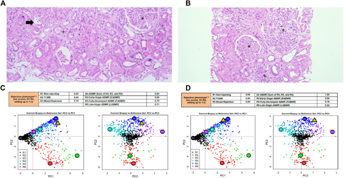
FIGURE 2.
Kidney graft histopathology and molecular microscope diagnostic report reports in patient #1 and patient #2. Kidney graft histopathology 12 months after application of MSCs in patient #1 (A) and patient #2 (B), hematoxylin eosin, x100. Molecular microscope diagnostic report (MMDx) for kidney transplant biopsy in patient #1 (C) and in patient #2 (D) 12 months after application of MSCs showing fully-developed antibody-mediated rejection with severe microvascular inflammation and molecular features of extensive interstitial fibrosis/tubular atrophy. *glomerulitis with transplant glomerulopathy (double contour formation), +interstitial fibrosis and tubular atrophy, → peritubular capillaritis.

