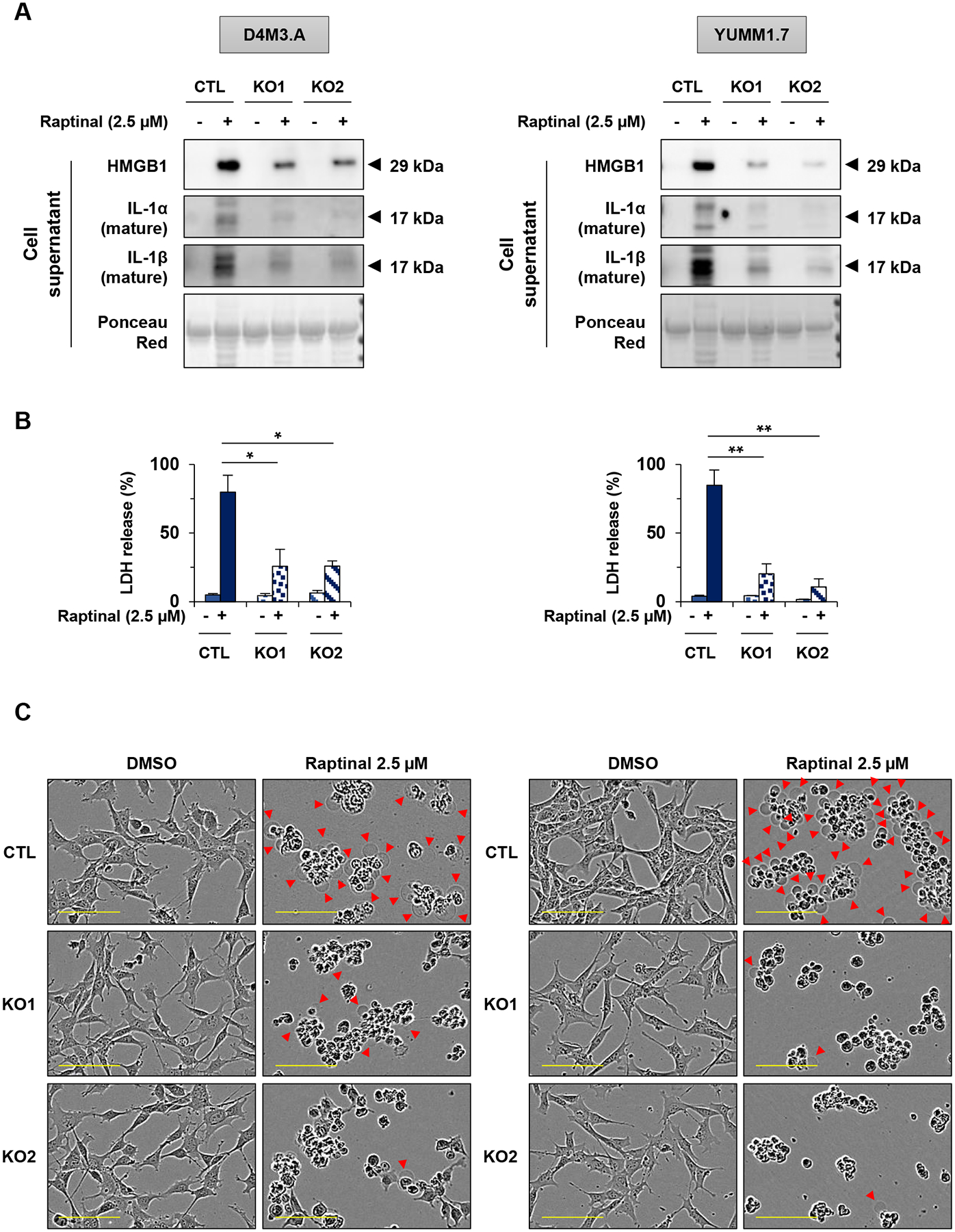
Figure 4: Raptinal-induced pyroptosis is GSDME-dependent in mouse melanoma cells.

(A-C) D4M3.A and YUMM1.7 mouse melanoma cells were treated with 2.5 μM of raptinal for 3 hours (A) Levels of secreted HMGB1, IL-1α and IL-1β in supernatants from wild-type (noted CTL) and two clones of D4M3.A or YUMM1.7 GSDME knockout cells (noted KO1 and KO2). Ponceau red as protein loading. Western Blots shown are representative of three independent experiments. (B) Percentage of LDH release averaged from three independent experiments. Error bars are SEM. Significance was assessed by Student’s t test, *p<0.05, **p<0.01. (C) Cell morphology visualized with the IncuCyte S3 system at x10 objective. Plasma membrane bubble-like protrusions, a characteristic feature of pyroptosis, are indicated by red arrow. Scale bar: 100 μm. Cell morphology images shown are representative of at least three independent experiments.
