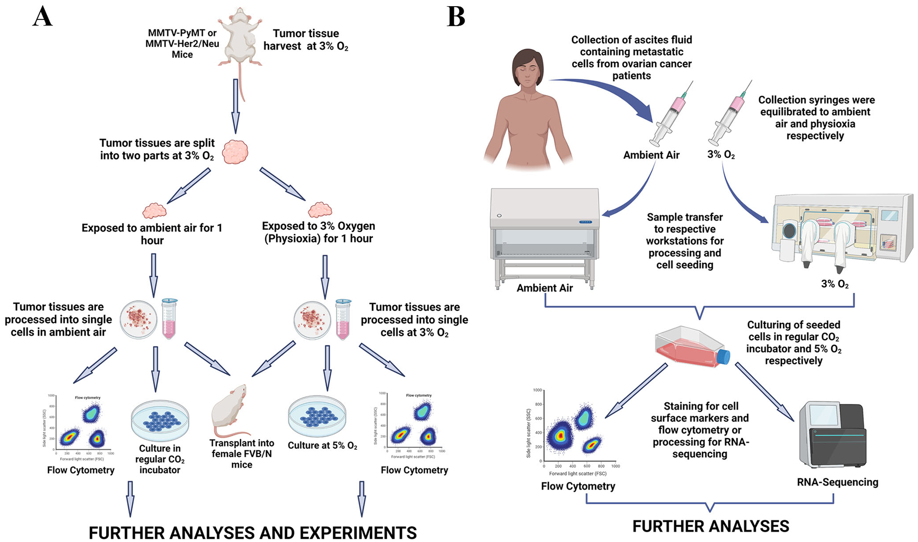
Figure 1.

Schematic representation of experimental workflow. A. Mouse mammary tumor tissues were harvested under physioxia, split into two parts and processed into single cells at respective oxygen tensions. Processed cells were either stained for flow cytometry, cultured for 5 days or transplanted into female FVB/N mice for further studies. B. Ascites fluid was collected from ovarian cancer patients with uniquely designed syringes previously equilibrated to ambient air and physioxia. Collected samples were transferred immediately to respective workstations, processed and cultured for 5 days prior to flow cytometric and sequencing analyses. Figures were created with BioRender.com.
