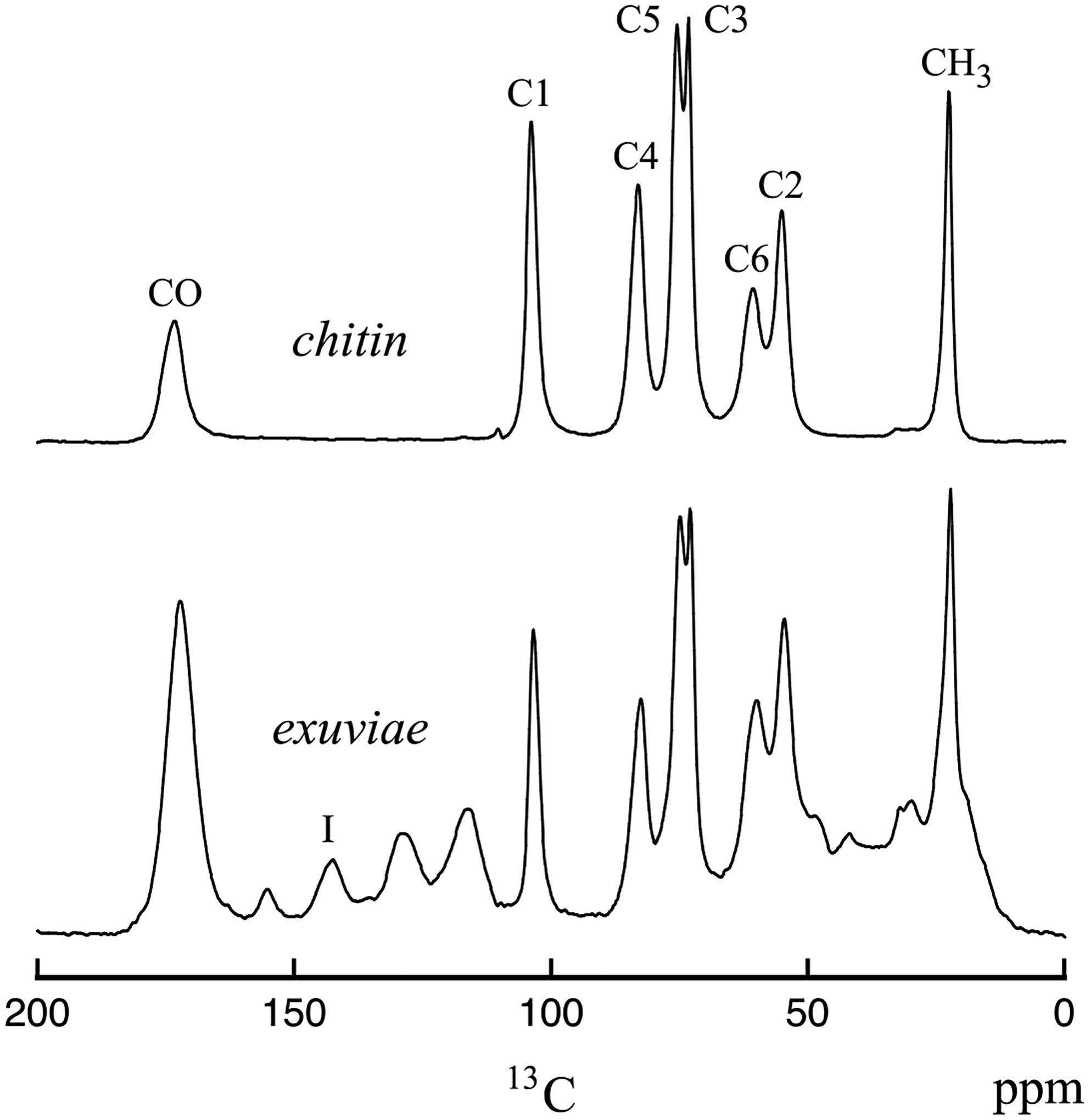
Figure 1.

13C CPMAS spectra of cicada exuviae (bottom) and chitin obtained from cicada exuviae (top). The spectra were obtained at room temperature on the 151 MHz (proton) spectrometer. The resonances shown in the chitin spectrum are labeled according to the chitin carbon positions for the structure shown in Table 1. The resonance labeled I in the exuviae spectrum arises from catechols.
