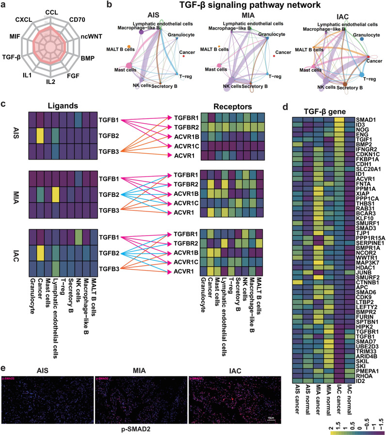
Fig. 5. Maps showing the role of the TGF-β signaling pathway in the dialog between cancer cells and the TME.
a Radar chart from CellChat data showing the major signaling pathways that mediate cell-to-cell interactions in LUAD. b TGF-β signaling pathway interactions between cancer cells and nine specific cell types of the TME from scRNA-seq data. The maps show TGF-β pathway interactions between cancer cells and cells of the immune microenvironment in AIS (left panel), MIA (middle panel), and IAC (right panel). c Heatmaps showing the distributions of ligands and receptors in the TGF-β pathway in the three stages of LUAD (upper: AIS, middle: MIA, lower: IAC). d GSVA of scRNA-seq showing that differences in the expression of genes (54 genes) downstream of the TGF-β pathway between normal cells and cancer cells differed among the three stages of LUAD. e TMAs were used to verify the expression of a gene related to TGF-β phosphorylation (p-Smad2) in the three stages of LUAD.

