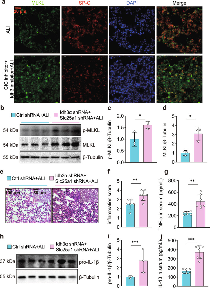
Fig. 5. Citratemt accumulation amplifies necroptosis and exacerbates LPS-induced lung tissue injury.
The mice were simultaneously injected with inhibitors of CIC and IDH3 or with sterile saline three days prior to intratracheal instillation of LPS (2.5 mg/kg) and were sacrificed 12 h after LPS administration. a Immunofluorescence staining and confocal microscopy were used to determine the localization of MLKL and SP-C. b–d Adenovirus vectors to specifically silence Slc25a1 and Idh3α in AECs were injected 10 days prior to intratracheal instillation of LPS (2.5 mg/kg), and the mice were sacrificed 12 h after LPS administration. MLKL and p-MLKL were detected by immunoblotting (n = 3). e Left lung tissue was embedded with paraffin and stained with hematoxylin and eosin (×100 and ×200 magnification). f The inflammation score was measured independently by three pathologists who were blinded to the experiment (n = 6–7). g Serum TNF-α levels were measured by ELISA (n = 6–7). h, i The expression of pro-IL-1β in the lungs was assessed by immunoblotting (n = 3). j Serum IL-1β levels were measured by ELISA (n = 6–7). The data are shown as the mean ± SD. *P < 0.05, **P < 0.01, and ***P < 0.001.

