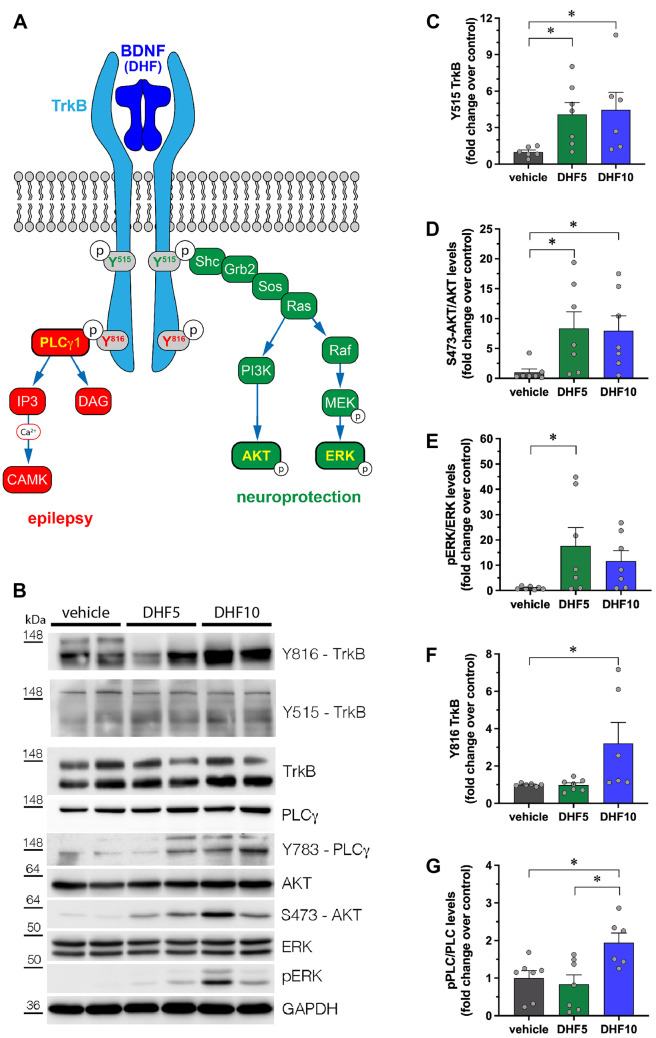
Fig. 5.
Phosphorylation of TrkB, AKT, ERK, and PLCγ proteins in the hippocampi of 7,8-DHF-treated rats. (A) Schematic representation of the different signaling pathways activated by BDNF (or 7,8-DHF) upon binding to TrkB receptor. (B) Representative western blot of the indicated proteins in extracts from hippocampi of DHF-treated rats. (C–G) Quantification of Y515 TrkB (C), S473-AKT (D), ERK (E), Y816 TrkB (F), and Y783-PLCγ (G) phosphorylation. Protein levels are shown as fold change over control (vehicle-treated rats). Levels of phosphorylated proteins are normalized against the corresponding total protein, then for loading (GAPDH). Vehicle: n = 7; 7,8-DHF 5 mg/kg: n = 7; 7,8-DHF 10 mg/kg: n = 7. *p < 0.05, Kruskal–Wallis one-way ANOVA, and post hoc Tukey’s test

