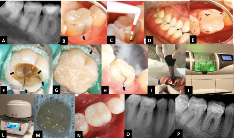
Figure 1.
Immediate treatment sequence: A) Initial digital radiography (DR) of tooth #46; B) Photo of the tooth, showing a vertical fracture line in the lingual surface of the tooth (BLUE ARROW); C) No periodontal probing depth was observed; D, E) Occlusal interferences in the working movements were investigated; F) Restoration in resin composite without cuspal coverage was removed, and several cracked lines in different directions under DOM (ARROWS) were identified; G) Temporary restoration in resin composite; H) Preparation of full crown of tooth #46; I) Intraoral CAD/CAM scanner; J) CEREC milling machine; L) The oven used for the preparation of the final restoration; M) Full crown in E-MAX; N) Definitive full crown restauration was placed; O) Periapical and bitewing radiography after 6 months; P) 5-year follow-up

