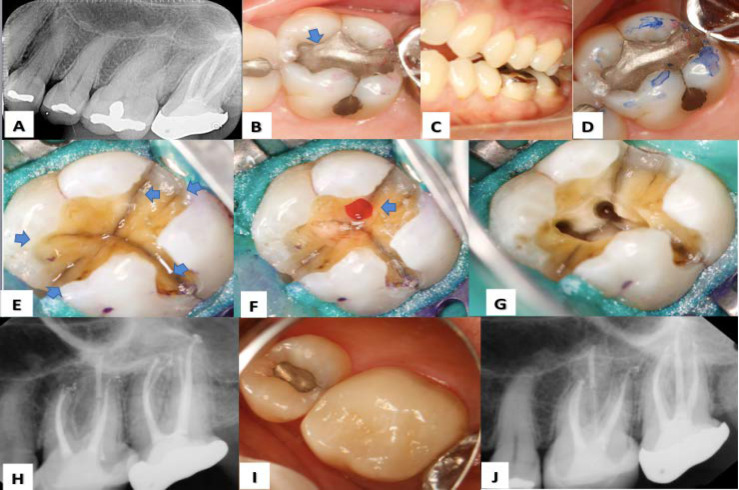
Figure 2.
Treatment sequence: A) Diagnostic radiograph of tooth #26; B) Clinical evaluation; identifying the presence of a class I amalgam restoration (BLUE ARROW); C, D) Occlusion evaluation; showing occlusal interferences in the lateral movement; E) Identification of crack lines after the removal of amalgam (BLUE ARROWS); F) Small direct pulpal exposure (BLUE ARROW); G, H) Endodontic treatment and final restoration by CAD-CAM System; I, J) Clinical and radiographic follow-up at 5 years

