
Extended Data Fig. 7 |. Loss of p53 in OCSCC induces adrenergic switch proximally in TG neurons.
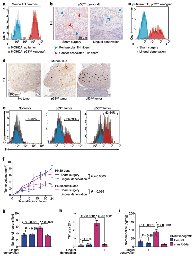
a, Flow cytometry quantification of neurotrophin-3-positive (NT3+), TH+ neurons in freshly collected ipsilateral TG neurons 3 weeks after orthotopic inoculation of p53null PCI-13 cells to the tongues of sympathectomized mice. Non-tumour-bearing, sympathectomized mice were used as controls (n = 6). b, Representative immunohistochemical analysis for TH+ in orthotopic xenografts; data independently replicated in 16 mice. c, Flow cytometry quantification of NT3+TH+ neurons in ipsilateral TG neurons (n = 6 mice per condition). d, Representative images of TH+ TG neurons in mice without tumours (left) and 3 weeks after injection of p53WT (middle) or p53null (right) PCI-13 cells to the ipsilateral tongue; data independently replicated in nine mice. e, Flow cytometry quantification of NT3+ TH+ neurons in freshly collected ipsilateral TG 3 weeks after orthotopic inoculation of p53WT (middle) and p53null (right) PCI-13 cells to the tongue. Non-tumour-bearing mice were used as controls (left, n = 12 per group). f, Serial in vivo analyses of tumour growth after engraftment of HN30 transfected with either control lentivirus (HN30-lenti) or shmiR34a (HN30-shmiR34a) into BALB/c (nu/nu) mice. Mice were randomized and underwent lingual denervation or sham surgery 1 week before cell injection (n = 8 per group). Tumour growth curves represent mean tumour volume ± s.e.m.; unpaired two-tailed t-test. g–i, Neural density (g), TH+ area (h), and noradrenaline levels in vivo (i) in HN30-lenti and HN30-shmiR34a orthotopic xenografts with and without lingual denervation (n = 5 biologically independent samples per condition). Bars indicate mean ± s.e.m.; unpaired two-tailed t-test.