Figure 1.
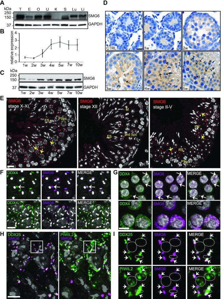
SMG6 localizes to the CB in round spermatids. (A) Western blotting of different tissues using anti-SMG6 antibody with anti-GAPDH as loading control. T, testis; E, epididymis; O, ovary; U, uterus; K, kidney; S, spleen; Lu, lung; Li, liver. (B) Relative expression of SMG6 during the first wave of spermatogenesis. Testis samples were collected from juvenile mice at different time points (1, 2, 3, 4 and 5 weeks) and adult mice (7 and 10 weeks). Two independent sample sets were included. Anti-SMG6 western blots were quantified with ImageJ software using anti-GAPDH for normalization. Error bars represent mean ± standard deviation (SD). (C) Representative western blot image of (B). (D) Immunohistochemistry of testis sections from 1, 2, 3, 4 and 5 weeks (w) old mice using anti-SMG6 antibody or no primary antibody (CTRL). Arrows point at selected examples of cytoplasmic SMG6 signal in round spermatids. Asterisks: elongating spermatid bundles. Scale bar: 5 μm. (E) IF of testis sections using anti-SMG6 antibody (red). PSpc, pachytene spermatocyte; RS, round spermatid; ES, elongating spermatid; M, meiotic metaphase plate; 2nd Spc, secondary spermatocyte. Arrows point at selected cytoplasmic granules. Scale bar: 10 μm. (F) Co-IF of SMG6 (magenta) and DDX4 (green) in round spermatids (upper panel, white arrow) and in late pachytene spermatocytes (lower panel, dashed circle). Scale bar: 10 μm. (G) Co-localization of SMG6 (magenta) with DDX4 (green) in human round spermatids (upper panel) and spermatocytes (lower panel). Arrows point at selected CBs. Scale bar: 10 μm. (H) SMG6 co-localizes with DDX25 and PIWIL2 to CB precursors in late pachytene spermatocytes (stage XI). SMG6 in magenta, DDX25 (left panel) and PIWIL2 (right panel) in green. Confocal 4-channel imaging was used to detect all three specific antibodies and DAPI in the same slides and artificial colors to each channel rendered afterwards using Adobe Photoshop. Scale bar: 10 μm. (I) The area indicated by a white box in panel H with higher magnification. Upper panel: co-localization of SMG6 with CB-specific DDX25. Lower panel: partial co-localization of SMG6 with PIWIL2. Co-localization points are marked by arrows. White dashed lines indicate examples of PIWIL2-positive (but DDX25- and SMG6- negative) IMCs. See also Supplementary Figure S1.

