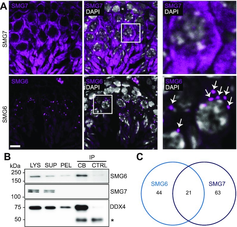
Figure 2.

Characterization of SMG6 and SMG7 during spermatogenesis. (A) IF of adult mouse testis sections with either anti-SMG6 or anti-SMG7 antibody (both in magenta). Scale bar: 10 μm. The areas indicated by white boxes are shown in higher magnification in the right panel. White arrows indicate perinuclear SMG6-positive granules in spermatocytes. (B) Western blotting of isolated CBs using anti-SMG6 and anti-SMG7 antibodies. LYS, lysate of cross-linked cells; SUP, supernatant after low-speed centrifugation; PEL, CB-containing pellet fraction; CB-IP, CBs isolated by anti-DDX4 IP; CTRL, control IP using rabbit IgG. Anti-DDX4 Western blotting confirms the successful CB isolation. IgG heavy chain is indicated with an asterisk. Cross-linking causes a very weak background precipitation of SMG6 and DDX4 in the control IgG IP. (C) Venn diagram showing the unique SMG6 (44), unique SMG7 (63) and shared (21) interaction partners identified in the mass spectrometry analysis. See also Supplementary Figure S2 and Supplementary Table S1.
