Figure 4.
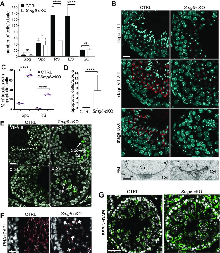
Spermatogenesis is arrested at round spermatid stage in Smg6-cKO mice. (A) Quantification of different cell types in CTRL and Smg6-cKO seminiferous epithelium. Quantified cell types included SOX3-positive spermatogonia (Spg), γH2AX-stained sex body-positive spermatocytes (Spc), round spermatids (RS) and elongating spermatids (ES) that were recognized on the basis of nuclear morphology and their position in the epithelium, and SOX9-positive Sertoli cells (SC). Cells were counted from a minimum of 70 tubules across three biological replicates per genotype. Error bars represent mean ± SD, and p-value from Mann-Whitney U-test, *P < 0.05, ****P < 0.0001, NS = non-significant. (B) PFA-fixed testis sections were stained with Rhodamine-conjugated PNA (red) to visualize acrosome and DAPI (blue) to visualize chromatin. Scale bar: 20 μm. The lower panel shows electron microscopy of the acrosomal region of round spermatids in control and Smg6-cKO mice. Nu, nucleus; Cyt, cytoplasm. Stars indicate the acrosome, which is fragmented into three separate granules in cKO. Arrows indicate the borders of the acrosomal region. Scale bar: 1 μm. (C) The percentage of tubules containing at least one apoptotic spermatocyte or round spermatids in a whole testis cross-section. At least 90 tubule cross-sections from each of the 3 CTRL and 3 Smg6-cKO mice were analyzed. Two-sided Fisher's exact test was used to demonstrate a significant association between CTRL and Smg6-cKO mice with the percentage of tubules with apoptotic cells, ****P-value < 0.0001. (D) Quantification of apoptotic cells per seminiferous tubule cross-section in CTRL and Smg6-cKO testes. Error bars represent mean ± SD, and P-value from Mann–Whitney U-test, ****P < 0.0001. (E) Representative TUNEL staining images of apoptotic spermatocytes (upper panel, stage VII-VIII) and round spermatids (lower panel, stage X–XI) on Smg6-cKO testis sections, together with matched stages from control testes without apoptotic cells. Scale bar: 20 μm. (F) PFA-fixed epididymides of control and Smg6-cKO mice were stained with PNA (red) and DAPI (white). Selected round spermatids with positive acrosome staining indicated by white arrows. Scale bar: 20 μm. (G) Testis sections were immunostained with anti-ESPIN antibody (green) to visualize apical ectoplasmic specializations at stage VIII-IX of the seminiferous epithelial cycle. Scale bar: 50 μm. See also Supplementary Figure S4.

