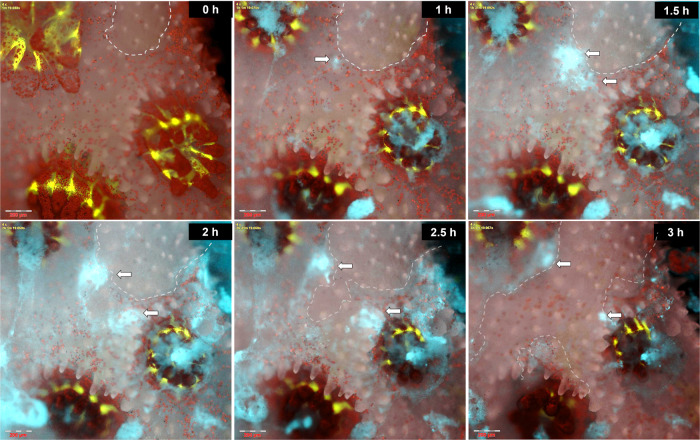
Fig. 3. Tissue lesions allow rapid colonization and infection by V. coralliilyticus.
A time series displaying V. coralliilyticus accumulation at the edge of a minor tissue lesion, with subsequent bacterial proliferation and extensive tissue necrosis. Time ‘0’ h–P. damicornis fragment immediately prior to inoculation. Coral GFP (green) and algal chlorophyll (red) are shown on the background of the colony’s skeleton (grey). Dashed line marks the borders of a small lesion approximately 300 μm in diameter. 1 h–V. coralliilyticus cells (cyan) settling at the lesion edge (arrow), are clearly distinct from pathogen-lades mucus flocs near polyp mouths. 1.5 h–3 h–further V. coralliilyticus accumulation at the torn tissue edge is followed by rapid lesion expansion and death of neighboring polyps. The complete sequence from this experiment is provided in Video S3. Scale bars are 200 μm.

