Figure 5.
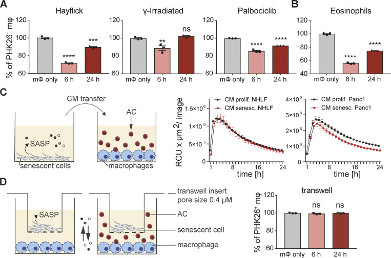
Senescent cells inhibit corpses removal through direct cell-cell contact. (A) Quantification of efferocytosis of apoptotic corpses by BMDMs in the presence of different types of murine 3T3 senescent cells (senescence induction by Hayflick limit, γ-irradiation or Palbociclib). BMDMs were primed in co-culture for 6 or 24 h. Apoptotic corpses (Jurkat cells) were labeled with PKH26 and added to macrophages in a 5:1 ratio. Samples were analyzed by flow cytometry 1 h post corpse exposure. All values in are means ± SEM; **P < 0.01, ***P < 0.001, ****P < 0.0001. Statistically significant differences were determined by one-way ANOVA with Bonferroni correction; n = 3 biological replicates. (B) Quantification of efferocytosis of apoptotic eosinophils by BMDMs in the presence of senescent 3T3 cells (induced by Palbociclib). All values in are means ± SEM; ****P < 0.0001. Statistically significant differences were determined by one-way ANOVA with Bonferroni correction; n = 3 biological replicates. (C) Conditioned media (CM) derived from either proliferating (black line) or senescent cells (red line) was transferred to naïve human MDMs. pHrodo labeled apoptotic corpses (AC, Raji cells) were added to MDMs and efferocytosis was monitored over time using the IncuCyte S3 System. Data are representative of three independent experiments. All values are means ± SEM. (D) Senescent cells (3T3, Palbociclib) were seeded in a transwell insert, which was placed into a well containing BMDMs. Efferocytosis of apoptotic corpses (Jurkat cells) by macrophages co-cultured with senescent cells in a transwell was analyzed by flow cytometry. Apoptotic corpses were labeled with PKH26 and added in a 5:1 ratio to the part of the well containing BMDMs. All values are means ± SEM. Statistically significant differences were determined by one-way ANOVA with Bonferroni correction; n = 3 biological replicates.

