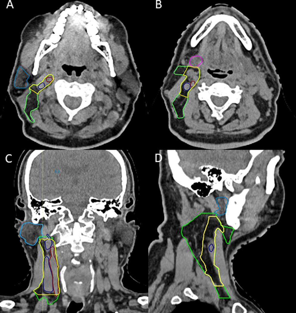
Figure 1.
CT neck slices in the axial (A and B), coronal (C), and sagittal (D) view demonstrating the differences between the modeled high-risk elective contralateral CTV (yellow) and the RTOG defined consensus elective neck contour (green) containing contours of the modeled CTV (green) and consensus CTV (yellow). The modeled high-risk CTV is defined by the extreme location of all LN centroids in each respective LN station. The internal jugular vein (dark blue), internal carotid artery (red), parotid gland (light blue), and submandibular gland (pink) are contoured for reference.

