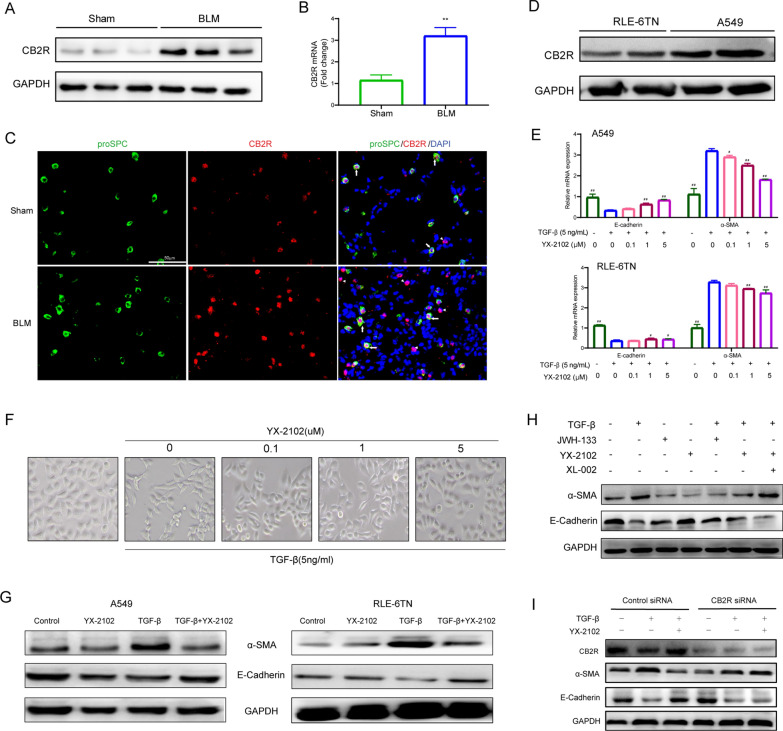
Fig. 4.
YX-2102 retarded TGF-β1-induced EMT in a CB2 R-dependent pathway. Rats were intratracheally treated with 5 mg/kg BLM once at day 0, followed by YX-2102 or vehicle treatment by intraperitoneal injection at a dose of 25 mg/kg daily. On day 21, rats were sacrificed and sampled. A, B Western blotting and qPCR were performed to evaluate the expression of CB2R in rats’ lung tissues of bleomycin-induced pulmonary fibrosis and sham group. C Immunofluorescence staining for Pro-SPC (green) and CB2R (red) in lung tissues. Arrows indicate epithelial cells with CB2R positive aggregates. Arrowheads indicate inflammatory cells with CB2R positive aggregates. Scale bar = 50 μm. D The expression of CB2R in A549 and RLE-6TN cells was evaluated by WB. E Cells were pretreated with 0.1, 1.0 or 5.0 µM indicated compounds for 2 h, followed by stimulation with 5 ng/mL TGF-β1 for another 48 h. DMSO treatment was set as control. The mRNA levels of E-cadherin and α-SMA were analyzed by qRT–PCR. F The morphological changes of A549 cells were observed at 48 h (magnification ×200). G Western blotting was performed to analyze the expression of E-cadherin and α-SMA in A549 (left panel) and RLE-6TN cells (right panel). H A549 cells were treated with YX-2102, JWH-133 or XL-002 for 2 h and stimulated with 5 ng/mL TGF-β1 for another 48 h. The levels of a-SMA and E-cadherin was assessed by western blotting. I Cells were transfected with 10 nM of control (scrambled) siRNA, CB2R-siRNA, subsequently pretreated with YX-2102 (5.0 µM) for 2 h, followed by TGF-β1 (5 ng/mL) treatment for 48 h. The expression of CB2R, α-SMA and E-cadherin was assessed by western blotting. Data are expressed as mean ± SEM, n = 5, **P < 0.01 versus the Sham group, #P < 0.05, ##P < 0.01 versus the TGF-β group (TGF-β = 5 ng/mL, YX-2102 = 0 µM)

