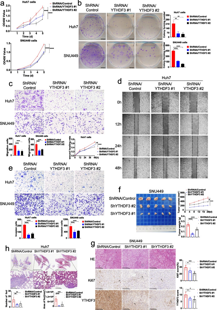
Fig. 3.
YTHDF3 knockdown inhibits proliferation, migration and invasion of Huh7 and SNU449 cells in vitro and tumor growth and lung metastasis of Huh7 and SNU449 cells in vivo. a-b CCK8 proliferation assays and colony formation assays were performed to determine cell proliferation of Huh7 and SNU449 cells after YTHDF3 knockdown. c-d Transwell migration assays and wound healing assays were performed to determine cell migration of Huh7 and SNU449 cells after YTHDF3 knockdown. The Image J software was used to quantified cells’ migration ability. Scale bar 200 μm. e Transwell invasion assays were performed to determine cell invasion of Huh7 and SNU449 cells after YTHDF3 knockdown. The Image J software was used to quantified cells’ invasion ability.Scale bar 200 μm. f Representative images of tumor growth in xenografted BALB/c nude mice with SNU449 knockdown YTHDF3 cells (left). The growth curves and the average weight of xenograft tumors were shown (right).g Representative images of HE and IHC staining of YTHDF3 and Ki67 of xenograft tumor (left), IHC scores of YTHDF3 and Ki67 were shown in bar graphs (right). Scale bar 200 μm. h Representative images of HE staining of orthotopic lung metastasis model (upper). The number and total area of metastatic foci were shown in bar graphs (lower). Scale bar 5 mm. Data are presented as mean ± SD (*p < 0.05, **p < 0.01, ***p < 0.001 and ****p < 0.0001). Arrows indicate the location of positive signal of YTHDF3 or Ki67 protein

