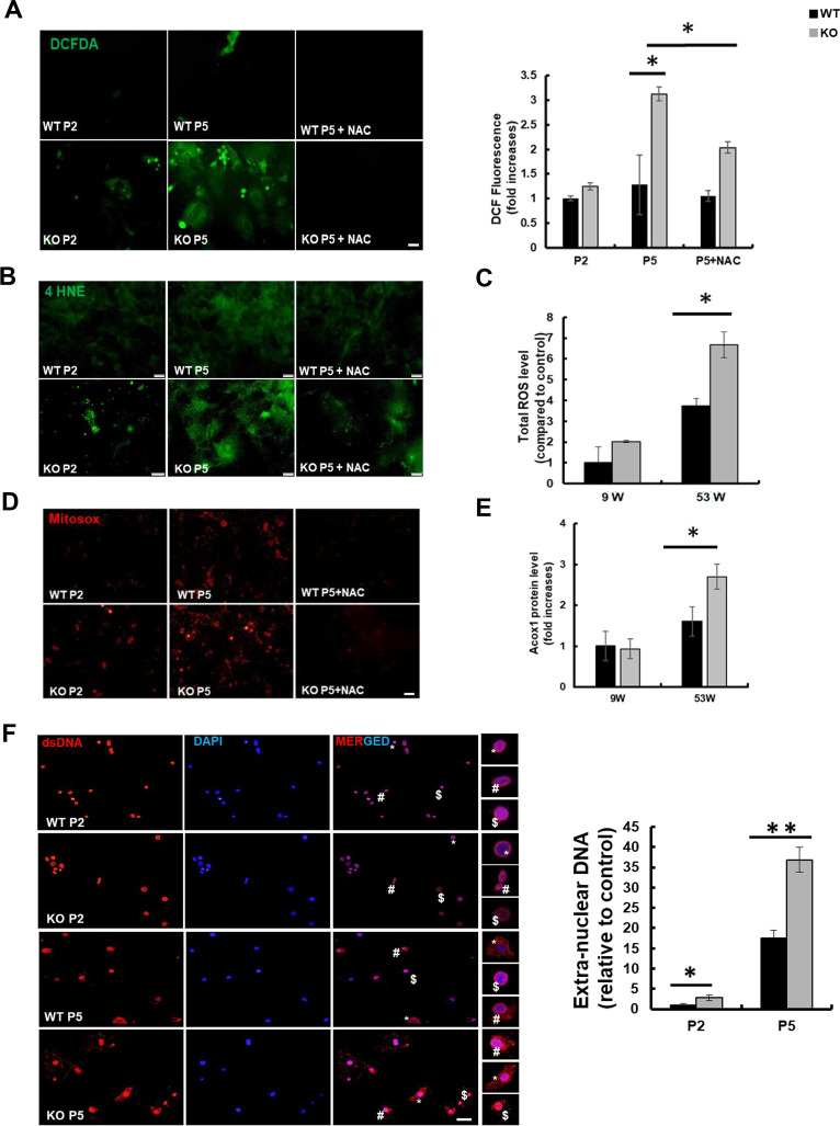
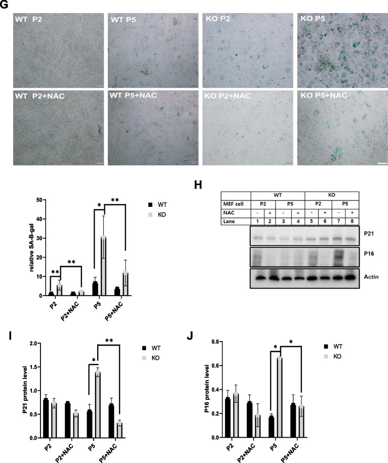
Fig. 2.
Aging is induced in catalase-deficient mice through ROS generation. A Representative fluorescence images of DCFH-DA staining of WT and catalase-KO MEFs at P2 and P5 levels. Quantification of cells showing green fluorescence (corresponding to DCFH-DA) and fluorescence intensity of MEFs. *P < 0.05 WT P5 versus KO P5; KO P5 versus KO P5 + NAC. B Representative fluorescence images of MEFs fixed and immunostained with anti-4HNE (green). Scale bar represents 20 μm. C Total ROS was measured in liver lysates of WT and catalase-KO mice at 9 W and 53 W. Values represent mean ± SD (n = 3, 4). *P < 0.05 WT 53 W versus KO 53 W. D Representative (red) fluorescence image of MitoSOX staining in MEFs, as in A. E ACOX1 levels were measured using the ACOX1 ELISA kit from the liver lysates of mice as in C. F IF staining of anti-dsDNA (red), DAPI (blue) in MEFs indicated as in A. Insets(*,#, $) enlarged cells; scale bar, 20 μm. Intensities of dsDNA positive cells at each passage, P2 and P5, in WT and KO MEFs.G MEFs at P2 and P5 were treated with 5 mM NAC overnight, and senescence-associated β-galactosidase staining was analyzed. The percentage of senescent cells was analyzed in WT and KO MEFs treated with NAC. Positive intensities of β-galactosidase staining were measured using the ImageJ software. Bar graph represents mean ± SD (n = 3 experiments). Scale bar represents 100 μm. *P < 0.05, WT P2 versus KO P2; KO P2 versus KO P2 + NAC; WT P5 versus KO P5. KO P5 versus KO P5 + NAC. H Proteins were extracted from MEFs as in G. Immunoblot analysis was performed using whole-cell lysates with indicated senescence-associated antibodies. I, J quantified protein level of P21 and P16 normalized with actin. Bar graph represents mean ± SD (n = 3 experiments). *P < 0.05; **P < 0.0001


