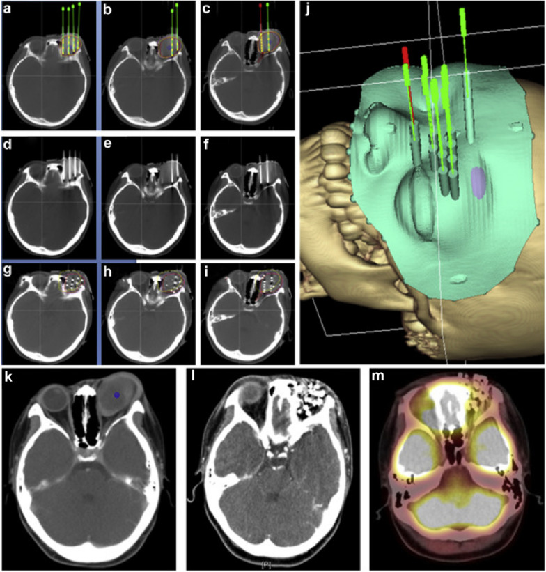
Fig. 2.
The CT-guided 125I seed implantation procedure and therapeutic efficacy. Figure a–c showed the preoperative treatment plan, including the planned needle locations, seed distribution, target volume doses, and organs at risk in a case of locally recurrent embryonal rhabdomyosarcoma of the orbit after surgery and EBRT. The green or red needles and yellow seeds were the simulated needles and seeds in the brachytherapy treatment planning system. Figure d–f showed the actual locations of the needles before the seed implantation. Figure g–i showed the actual distribution of seeds and the doses in target volume and organs at risk after seed implantation. Figure j showed the 3D-printed non-coplanar template model with guide holes on it in brachytherapy treatment planning system. Figure k–m showed CT or PET-CT images of the tumor in preoperation, 3-month postoperation, and 6-month postoperation, and PET-CT showed no residual tumor with metabolic activity in Figure m [41]

