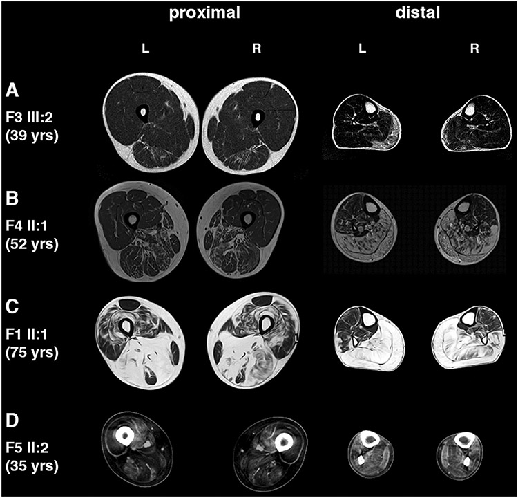
Fig. 2.
Muscle imaging. (A) Patient F3 III:2 at age 39. Moderate fatty degenerate changes are seen in gastrocnemius medialis muscle more on the right and mild in semitendinosus muscle. (B) Patient F4 II:1 at age 52. Severe involvement in all calf muscles and milder in hamstrings. (C) Patient F1 II:1 at age 75. Severe changes are seen in all calf muscles symmetrically and peroneus longus on the left. Hamstring muscles are also severely affected with less involvement in vastii muscles. Rectus femoris is best spared muscle. (D) Patient F5 II:2 at age 35. Severe involvement in anterior and posterior thigh muscles and diffuse fatty degenerative changes in all lower leg muscles, most prominent in peroneus longus and tibialis posterior.

