-
A
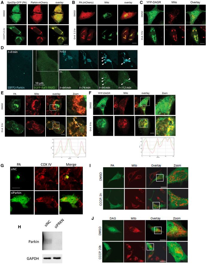
Hela cells were transfected with the Spo20p‐GFP PA reporter and mCherry‐Parkin followed by CCCP treatment for 5.5 h. Note that CCCP treatment led to the recruitment of Spo20p‐GFP to Parkin‐positive mitochondria. Scale bar = 10 μM.
-
B, C
HeLa cells were transfected with Raf1‐PABD‐mCherry PA reporter or YFP‐DAGR with Parkin‐FLAG followed by antimycin (4 μM) and oligomycin (10 μM; O + A) treatment to activate mitophagy. Note that Raf1‐PABD‐mCherry PA reporter (B) and YFP‐DAGR (C) both translocated to mitochondria (TOM20) after the treatment. Scale bar = 10 μM.
-
D
Hela cells expressing KR‐dMito and EBFP2‐Parkin (cyan) were illuminated at 559 nm to induce mitophagy. Images were acquired at indicated time points after illumination. Note that PA accumulation on Parkin‐positive mitochondria is induced by photodamage (t = 94 min and 112 min). Arrowheads point to the co‐accumulation of Parkin and the PA reporter on damaged mitochondria. Scale bar = 10 μM.
-
E, F
SH‐SY5Y cells were transfected with (E) the Raf1‐PABD‐GFP PA reporter or (F) YFP‐DAGR followed by antimycin (4 μM) and oligomycin (10 μM) or DMSO treatment, as indicated. Both reporters became enriched in the mitochondria (TOM20). Line scan analysis (Image J software) under the images indicates colocalization between the lipid reporters (green) and mitochondria (red) corresponding to the lines drawn in the images. Scale bar = 10 μm.
-
G
Sh‐Sy5Y cells were transfected with control (siNC) or Parkin siRNA (siParkin) followed by the PA‐GFP reporter and treated with CCCP (5.5 h). Note that Parkin KD prevented mitochondrial (COX IV) aggregation and PA concentration. Scale bar = 10 μM.
-
H
Immunoblotting confirmed Parkin KD's efficiency.
-
I, J
Hela cells were transfected with PA reporter and DAGR‐YFP without Parkin, by CCCP treatment, as indicated. Note that there is no accumulation of PA and DAGR reporters on mitochondria after CCCP treatment (by a Tom20 antibody). Scale bar = 10 μM.

