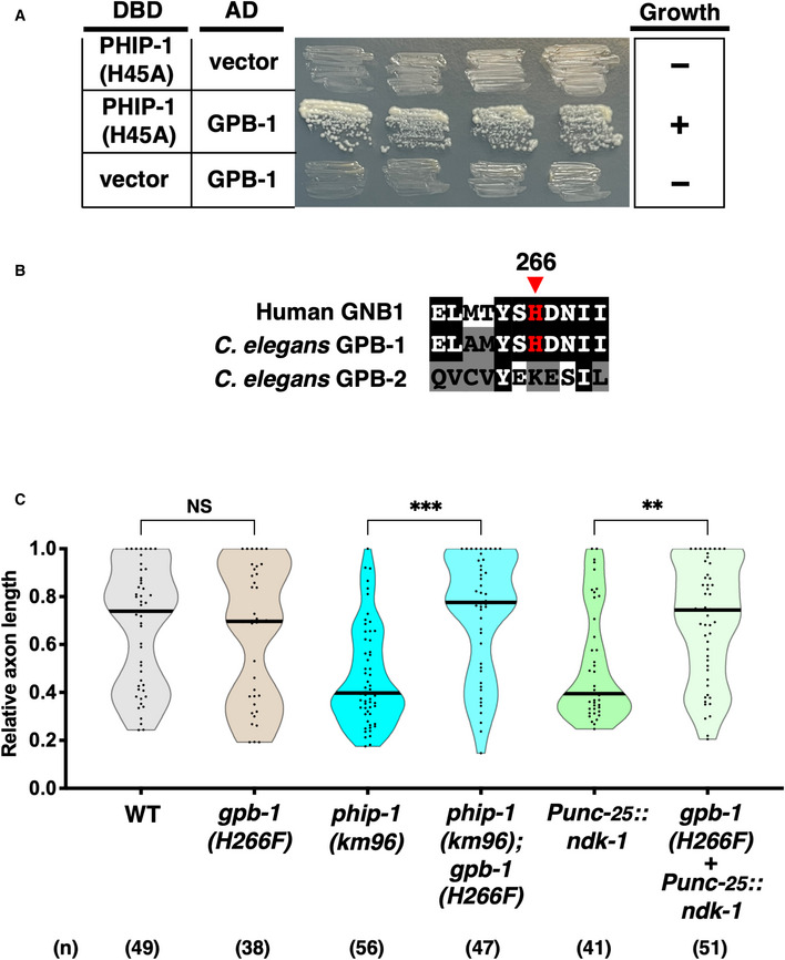
Figure 3. NDK‐1 and PHIP‐1 regulate axon regeneration through His‐phosphorylation of the Gβ subunit GPB‐1.

- PHIP‐1 interaction with GPB‐1 by yeast two‐hybrid assay. The reporter strain PJ69‐4A was co‐transformed with expression vectors encoding GAL4 DBD‐PHIP‐1(H45A) and GAL4 AD‐GPB‐1, as indicated. Yeast strains carrying the indicated plasmids were cultured on a selective plate lacking histidine and containing 5 mM 5‐aminotriazole for 4 days.
- His‐phosphorylation site in Gβ. Sequence alignments of the His‐phosphorylation site and flanking amino acids among human GNB1, GPB‐1, and GPB‐2 are shown. Identical and similar residues are highlighted with black and gray shading, respectively. The His‐phosphorylation site, His‐266, is indicated by an arrowhead.
- Relative axon length 24 h after laser surgery at the young adult stage. The number (n) of axons examined from three biological replicates is indicated. The black bar in each violin plot indicates the median. **P < 0.01, ***P < 0.001, as determined by the Kruskal–Wallis test and Dunn's multiple comparison test. NS, not significant.
