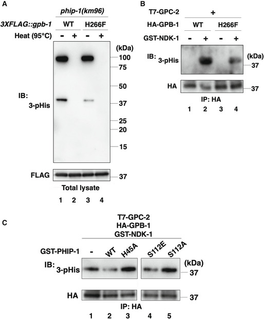
Figure 4. His‐phosphorylation of GPB‐1.

- His‐phosphorylation of GPB‐1 in animals. The phip‐1(km96) mutant animals carrying the 3XFLAG::gpb‐1 or 3XFLAG::gpb‐1(H266F) knock‐in allele were lysed. The lysates were treated with or without heating (95°C) and immunoblotted (IB) with anti‐3‐pHis and anti‐FLAG antibodies.
- NDK‐1 phosphorylates GPB‐1 in vitro. COS‐7 cells were co‐transfected with HA‐GPB‐1 or HA‐GPB‐1(H266F) and T7‐GPC‐2, and cell lysates were immunoprecipitated (IP) with anti‐HA antibodies. Immunopurified GPB‐1 was subjected to the in vitro kinase assay with recombinant GST‐NDK‐1. Phosphorylated GPB‐1 was detected by immunoblotting (IB) with anti‐3‐pHis antibodies.
- PHIP‐1 dephosphorylates GPB‐1 in vitro. COS‐7 cells were co‐transfected with HA‐GPB‐1 and T7‐GPC‐2, and cell lysates were immunoprecipitated (IP) with anti‐HA antibodies. The immunopurified HA‐GPB‐1 was first subjected to the in vitro kinase assay with recombinant GST‐NDK‐1. Phosphorylated GPB‐1 was then equally aliquoted and subjected to the in vitro phosphatase assay with recombinant GST‐PHIP‐1 or its variants. Phosphorylated GPB‐1 was detected by immunoblotting (IB) with anti‐3‐pHis antibodies.
