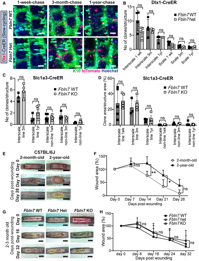
Figure EV4. Fbln7 knockout does not affect the maintenance of slow‐cycling stem cells or wound healing in young mice.

-
ADlx1CreER lineage tracing in the Fbln7 WT and Het backgrounds. Low‐dose tamoxifen was administered once at 2 months of age and samples were collected after 1‐week, 3‐month, and 1‐year chases. Wholemount staining of tail epidermis with tdTomato, K10, and Hoechst. Scale bar: 200 μm.
-
BThe number of Dlx1CreER clones in the scale or interscale (both line and non‐line) of Fbln7 WT mice for 1‐week (N = 4), 3‐month (N = 4), 1‐year chase (N = 3) and Fbln7 het mice for 1‐week (N = 5), 3‐month (N = 3), and 1‐year (N = 3) chase. ns, not significant. Mann–Whitney test.
-
CThe number of Slc1a3CreER clones in the interscale non‐line or interscale line regions for 1‐week, 3‐month or 1‐year chase. Fbln7 WT mice for 1‐week (N = 5), 3‐month (N = 3), and 1‐year chase (N = 3). Fbln7 KO mice for 1‐week (N = 5), 3‐month (N = 4), and 1‐year (N = 6) chase.
-
DQuantitation of the area of Slc1a3CreER clones per structural area from the same experiment as in (C).
-
E, FRepresentative pictures from tail wounds of 2‐month versus 2‐year‐old C57BL/6J wild‐type mice (E) and measurements of wound area over time (F). N = 6 (2‐month‐old) and N = 5 (2‐year‐old). Scale bar: 4 mm. **P < 0.01; *P < 0.05.
-
G, HRepresentative pictures from tail wound healing experiment in 2‐ to 3‐month‐old Fbln7 mice (G) and the wound area quantitation over time (H). N = 4 (WT), N = 8 (Het), and N = 7 (KO). Scale bar: 4 mm.
Data information: All graphs show mean SD. N reflects the number of biological replicates summarized from at least two independent experiments. Mann–Whitney test was performed for (B–D). two‐way ANOVA (Tukey's multiple comparison test) was performed for (F, H).
