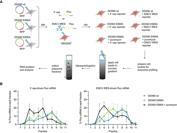
Figure 5. Polysome profile of type II IRES Fluc mRNA in the presence of DDX60.

- Schematic of polysome profiling strategy. HEK293T cells were transfected with DDX60 wt or DDX60 E890A (negative control). 48‐h post‐transfection, cells were transfected with in vitro transcribed 5′ cap‐ or EMCV IRES‐driven Fluc mRNA constructs. 16‐h post‐transfection, duplicate samples were treated with 200 μM puromycin for 20 min as positive controls for decrease in polysomes. Cells were treated with 100 μg/ml of cycloheximide for 15 min to arrest polysomes and subjected to polysome profiling by ultracentrifugation through 15–50% sucrose gradients. Amount of Fluc reporter mRNA from polysome fractions was determined by RT‐qPCR.
- Effect of DDX60 on 5′ cap (left) or EMCV IRES (right) driven Fluc mRNA polysomes.
Data information: Mean percent ± SEM of Fluc mRNA of each fraction relative to Fluc mRNA in all fractions, from n = 3 biological replicates. Full polysome profiles are shown in Fig EV5.
