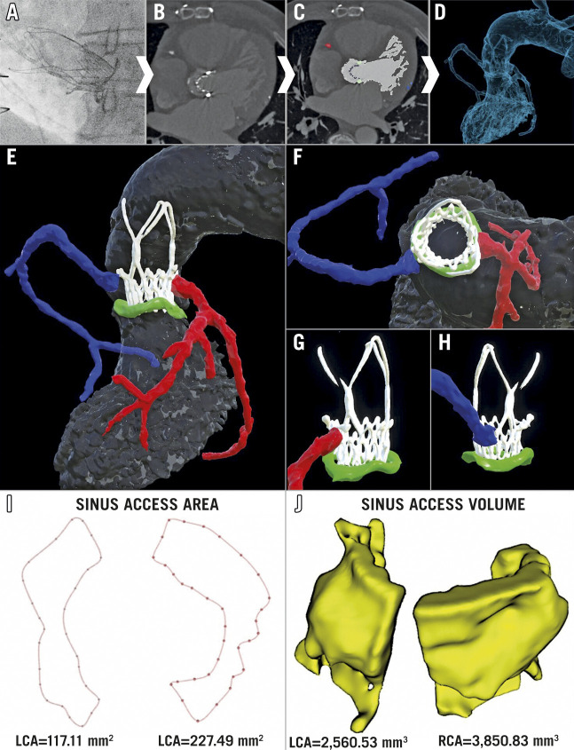
Figure 1. Multi-parametric 3D imaging evaluation of coronary access.
A post-procedural CT scan was performed and specific structures and regions of interest deemed relevant for evaluating coronary access were segmented to create a 3D digital model (A-D). Further image processing was then applied to improve the visualisation and the understanding of the geometrical relationships between the aortic sinuses (grey), ACURATE neo valve (white), surgical bioprosthesis (green), right coronary artery (blue), and left coronary artery (red) (E-H). In order to evaluate the complex 3D geometry of the sinus space, both (I) the size and morphology of the area available between the valve frame and the aortic wall to enter the sinuses (sinus access area), and (J) the volume available within the sinuses (sinus access volume) were determined for each ostium.

