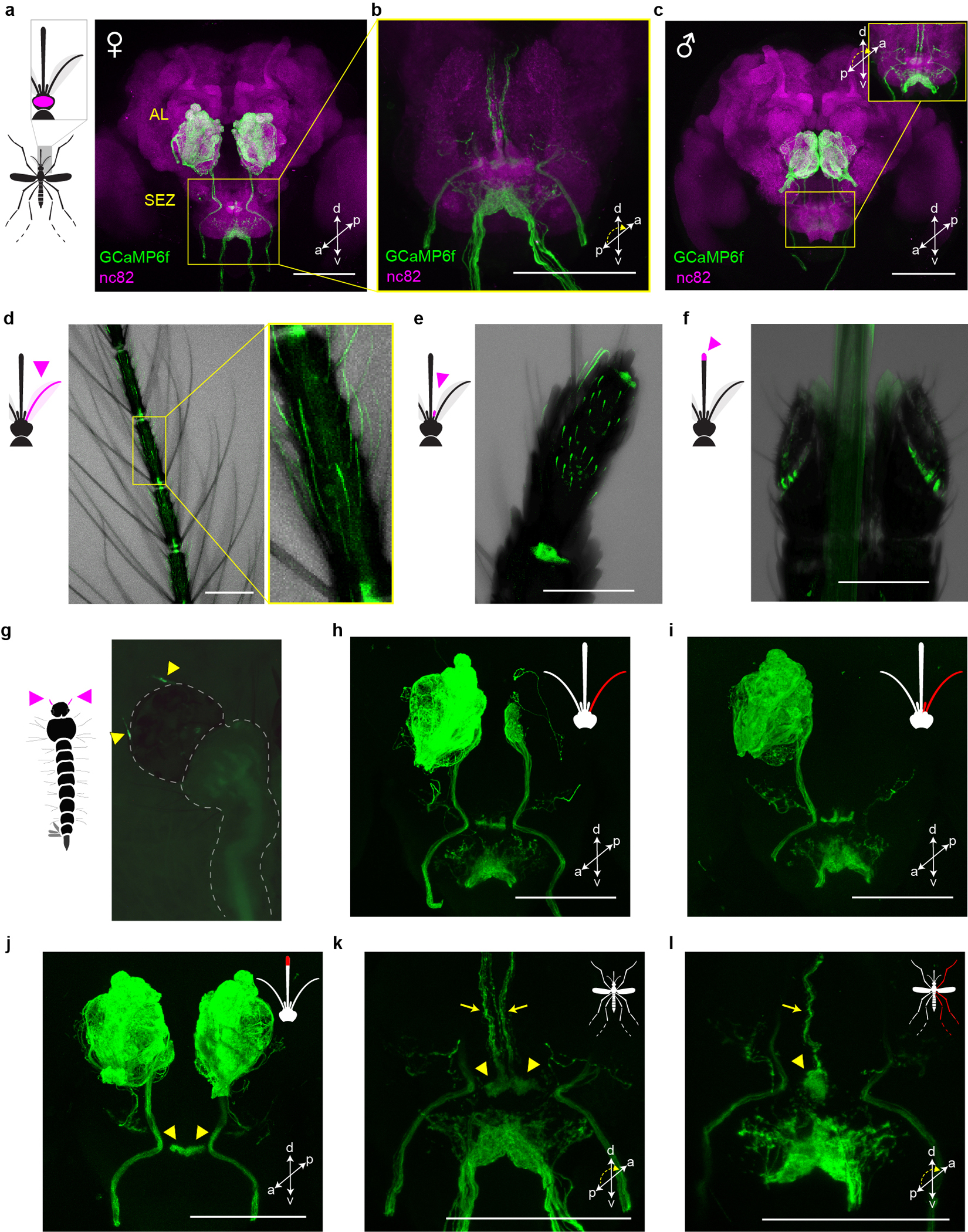
Extended Data Fig. 1 |. orco-T2A-QF2-QUAS-GCaMP6f labels chemosensory neurons in peripheral organs that project to the brain.

a–c, Antibody staining in female (a,b) and male (c) brains showing GCaMP in sensory neurons that innervate the antennal lobe (AL) and suboesophageal zone (SEZ). SEZ in (b) and the inset of (c) are viewed from posterior to better visualize GCaMP signal. d–g, Intrinsic GCaMP fluorescence in sensory neurons of adult female antenna (d), maxillary palp (e), labella (f) and larval antennae (g, arrowheads). Transmitted light image overlaid in (d–f). h–l, Antibody staining in brains of female mosquitoes with severed sensory organs (red in mosquito schematics). Severing right antenna only (h), or both right antenna and right palp (i) led to loss of signal in all ipsilateral glomeruli except two in the posterior-medial region or all ipsilateral glomeruli, respectively. We therefore infer that Orco+ AL glomeruli are innervated by sensory neurons in the ipsilateral antenna (n~32 glomeruli) and palp (n=2 glomeruli)51,52. Severing the tip of the proboscis (including the labella) led to loss of signal throughout the ventral SEZ (j), consistent with work in Anopheles gambiae indicating that Orco+ labellar neurons innervate this region31. However, labellum-less animals retained signal in an area of the dorsal SEZ recently termed the suboesophageal glomeruli (arrowheads in j) (https://www.mosquitobrains.org/). Signal in this dorsal region and corresponding ascending nerves was present in intact animals (k, arrowheads and arrows, respectively) but was lost when the ipsilateral legs were severed (l). This suggests that Ae. aegypti has Orco+ neurons on the legs that project to the SEZ, consistent with electrophysiological responses to olfactory stimuli on legs53. All scale bars 100 μm.
