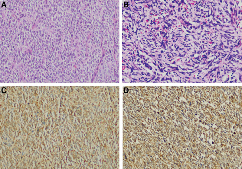
Figure 2.
The microscopic examination of the biopsy specimen (A: primary tumor, B: recurrent tumor) demonstrates a myxoid tumor with spindle- to oval-shaped cells with euchromatic nuclei and clear cytoplasm with arcuate vasculature. The tumor cells were immunohistochemically positive for Vimentin (C) and Bcl-2 (D).

