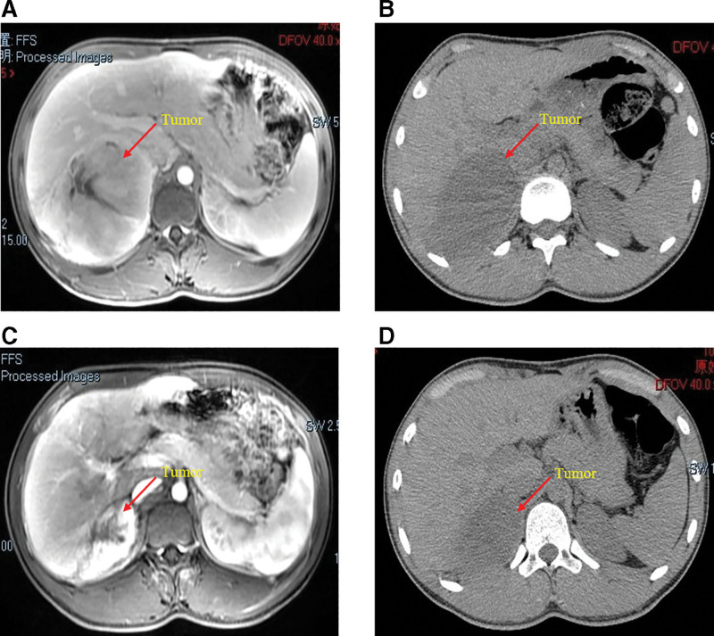
Figure 3.
The MRI (A) of the patient shows a recurrent mass in the operative area, which adhered closely to the right lobe of the liver. The CT scans show that the mass gradually shrinks after the second (B), fourth (C), and sixth cycles (D) of neoadjuvant therapy. CT = computed tomography, MRI = magnetic resonance imaging.

