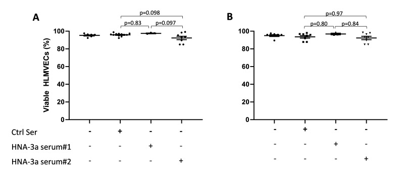
Figure 2. In vitro models of neutrophil-independent anti-HNA-3a-mediated endothelial cytotoxicity.
(A–B) Human lung microvascular endothelial cells (HLMVECs) were grown to 80–90% confluence in 12-well plates and left untreated (one hit model; A) or stimulated with 2 μg/mL lipopolysaccharide (LPS; two hit model; B) for 6 hours, followed by the addition of either buffer, control serum (Ctrl Ser) or anti-HNA-3a sera (anti-HNA-3a serum #1 or anti-HNA-3a serum #2). All sera were 10% final volume. After 30 min plates were flicked, cells were stained with Trypan blue and images were acquired. Viable HLMVECs were counted with ImageJ software. Data are presented as mean ± SE. p<0.05 was considered statistically significant.

