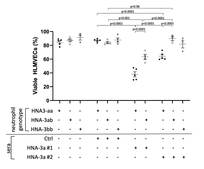
Figure 4. Two hit in vitro model of neutrophil-dependent anti-HNA-3-mediated endothelial cytotoxicity.
Human lung microvascular endothelial cells (HLMVECs) were grown to 80–90% confluence in 12-well plates and were stimulated with 2 μg/mL LPS for 6 hours, followed by the addition of buffer, freshly isolated HNA-3aa neutrophils (black dots), HNA-3ab neutrophils (white dots), or HNA-3bb neutrophils (grey triangles) that were allowed to settle for 30 min. Then cells were treated with either buffer, control serum (Ctrl serum) or anti HNA-3a sera (anti-HNA-3a serum#1 or anti-HNA-3a serum #2). All sera were 10% final volume. After 30 min plates were flicked, cells were stained with Trypan blue and 3–7 fields per well were acquired. Viable HLMVECs were counted with ImageJ software. Data are presented as mean ± SE. p<0.05 was considered statistically significant.

