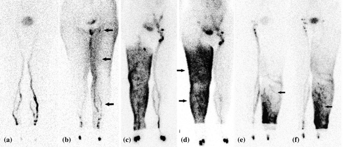
Fig. 3.
Lymphoscintigraphy imaging of the lymphatics. a, b Images of type II in a patient with left lymphedema a 30 and b 120 min after injection of contrast medium. Lymph stasis in the lymphatics (arrow) and visible dermal backflow (arrow) on the left thigh can be seen. The inguinal lymph nodes are reduced in number (arrow). c, d Images of type III in a patient with right lymphedema c 30 and d 120 min after injection of contrast medium. Dermal backflow (arrows) in the leg and thigh can be seen. e, f Images of type IV in a patient with left lymphedema e 30 and f 120 min after injection of contrast medium. Dermal backflow [arrow in e] and lymph stasis in the lymph vessels [arrow in f] in the leg can be seen and remains in the leg 120 min later. Reprinted from Maegawa et al.35 with permission from John Wiley & Sons, Inc

