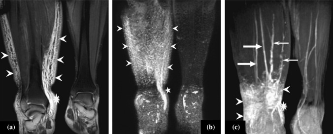
Fig. 5.
a Coronal T2-weighted 2D-TSE image with fat suppression shows an extensive reticular pattern of dilated lymphatic vessels, indicating neovascularization due to obstruction in the right lower leg (arrowheads). b Frontal 3D heavy T2-weighted MIP image demonstrates the same changes in the right lower leg (arrowheads). c Frontal 3D spoiled gradient-echo T1-weighted MRL MIP image obtained 35 min after Gd-BOPTA injection. Two slightly enlarged lymphatic vessels are visualized in the affected right lower leg (small arrows). The concomitantly enhanced veins (large arrows) show lower signal intensity. Furthermore, areas of accumulated lymph fluid are detected in the three modalities image (asterisk). No lymphedema is seen in the left lower leg. 2D-TSE two-dimensional turbo spin-echo, 3D three-dimensional, MRL magnetic resonance lymphography. Reprinted from Lu et al.162 with permission from Elsevier

