Fig. 1.
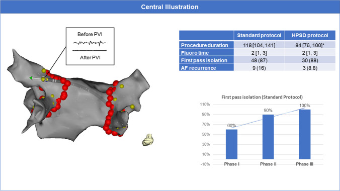
Efficacy and safety of a high-power short-duration ablation index–guided protocol for pulmonary vein isolation using a single catheter. Left: Documentation of entrance and exit block using a single catheter: After anatomical circumferential ablation around the pulmonary veins (red tags), the ablation catheter was positioned (with adequate contact force displayed by the orientation of the force vector) distal to the circumferential lesion to check for local signals. If no signals were identified (entrance block), pacing for exit block from the vein was performed at the anatomical ostium of every vein at four evenly distributed locations (yellow tags) in each pulmonary vein by pacing with 10 V and a pulse width of 1.5 ms. Right, top: Outcome measures for a control ablation protocol using a single catheter versus a high-power short-duration ablation index–guided protocol for PVI using a single catheter. * represents a p value of < 0.05. Right, bottom: To account for a potential learning effect during implementation of the single-catheter protocol, we arbitrarily split the control ablation protocol group into three consecutive groups (Phases I–III). Over time, the amount of first-pass isolation significantly increased (p = 0.01) from 60% after 11 pts to 100% after 41 cases

