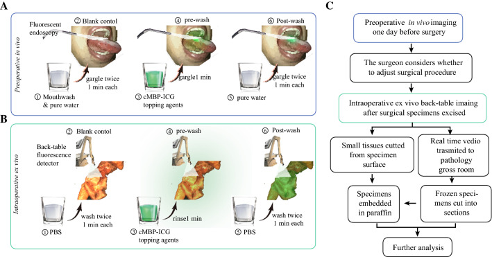
Fig. 1.
Workflow of fluorescence imaging-based head and neck squamous cell carcinoma (SCC) delineation. A After careful mouth-washing (step 1), patients (n = 10) with clinically suspicious or biopsy-proven head and neck SCC were examined using a Digital Precision Medicine imaging device H2000 (DPM, Beijing, China) with an endoscopic camera and an ICG-optimized Light Emitting Diode (LED)-filter system. The tumor area and the surrounding margins were imaged as a blank control (step 2). After gargling, a solution of cMBP-ICG (step 3) at different concentrations (2.5 μM or 5 μM, each dose n = 5) for 1 min, a pre-wash imaging procedure was performed (step 4). The patients then gargled ultrapure water for 1 min as a clearing solution (step 5), followed by cMBP-ICG administration of post-wash imaging (step 6). B Immediately after resection, tissue specimens, including margin samples, were imaged ex vivo in a vertical near-infrared imaging device Z2000 (DPM) following the same protocol described earlier. C Preoperative images were taken into consideration during the surgical procedure plan. Intraoperative back-table videos were transmitted in real time to the pathology gross room as frozen section’s references. Small tissues were cut from specimens’ high and low fluorescence intensity surfaces, respectively, for further analysis. cMBP-ICG, c-MET-binding peptide-indocyanine green

