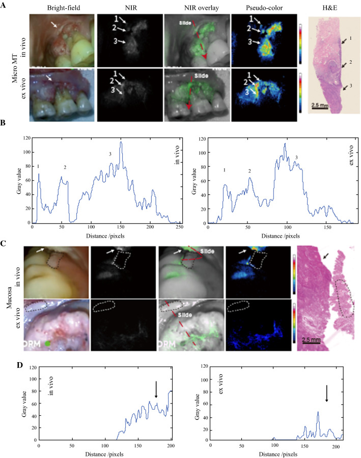
Fig. 3.
A Suspicious lesions discovered and C a nontumor lesion screened out during preoperative imaging procedure. Frozen sections were cut according to the real-time videos. The section locations (red dashed line) were ensured to meet the region of interest. A Severe dysplasia (arrows 1 and 2) detected 2 mm above the primary gum cancer area (arrow 3). B Two-dimensional (2D) fluorescence-intensity histogram illustrating the signal enhancement in the tumor area and the suspicious region. C A gum leukoplakia (dashed circle) adjacent to primary tongue cancer (arrow) that proved to be normal mucosa pathologically. D A 2D fluorescence-intensity histogram illustrating the signal enhancement in the tumor area. NIR, near infrared; H&E, hematoxylin and eosin

