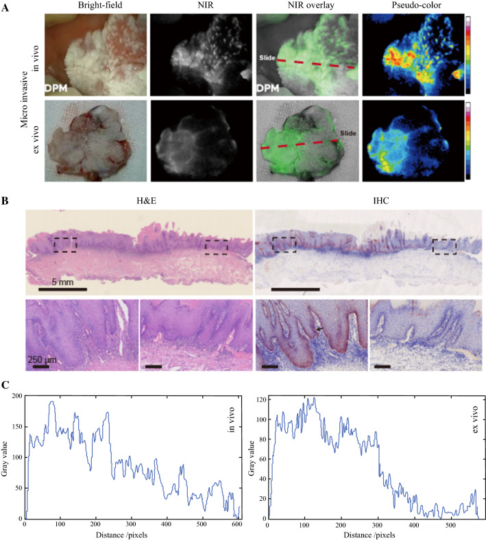
Fig. 4.
Heavily keratinized mucosa with small cancer foci in one patient. A Snapshots from preoperative (upper row) and intraoperative (lower row) imaging. B Hematoxylin and eosin (H&E) and immunohistochemistry (IHC) of the frozen sections. The lower row demonstrates enlarged views of the two areas framed in black in the upper row. The section locations (red dashed line in panel A) were ensured to meet the region of interest. C Two-dimensional (2D) fluorescence-intensity histogram illustrating the signal enhancement in the high c-Met expression area

