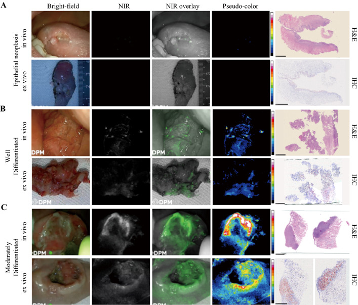
Fig. 5.
Comparison of epithelial neoplasia and two grades of head and neck squamous cell carcinoma (SCC). A Epithelial neoplasia showing low fluorescence intensity (FI) in both in vivo and ex vivo imaging. The frozen section shows low c-MET expression of this lesion. B Highly differentiated head and neck SCC showing slightly increased FI compared with panel A. The frozen section shows about 25% of the c-MET expression area fraction. C Moderately differentiated head and neck SCC showing high FI in in vivo and ex vivo imaging. The frozen section shows about 90% of the c-MET expression area fraction

