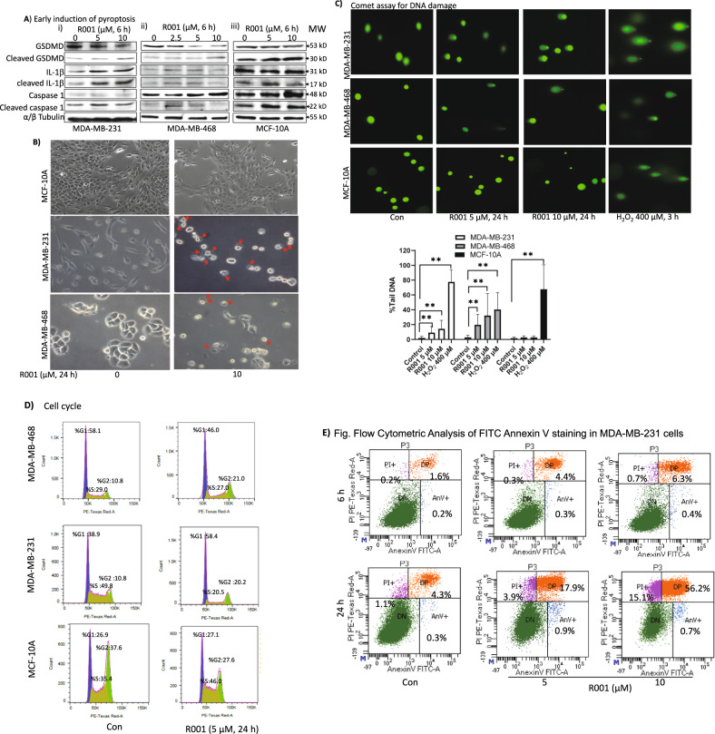
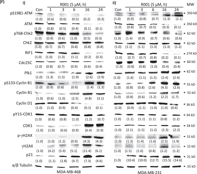
Fig. 5. R001 induces early pyroptosis and late DNA damage, altered cell cycle profile, and cell death in triple-negative breast cancer cells.
A–E Triple-negative breast cancer, MDA-MB-231 and MDA-MB-468 cells, or normal human breast epithelial cells, MCF-10A in culture were untreated or treated with 2.5, 5 or 10 µM R001 for 6 or 24 h and A whole-cell lysates were prepared and immunoblotted for gasdermin D (GSDMD), cleaved-GSDMD, interleukin-1β (IL-1β), cleaved IL-1β, caspase 1, and tubulin; B analyzed for morphology changes under phase-contrast microscopy imaging; red arrows show swollen cells; C 24 h subjected to Comet assay. Images were taken under a fluorescence microscope (upper) and quantified for the percent Tail DNA and plotted (lower); n = 3; D processed, and subjected to flow cytometry analysis for cell cycle progression; E cells were processed for Annexin V binding/flow cytometric analysis; and F MDA-MB-231 or MDA-MB-468 cells in culture were untreated (Con) or treated with 5 µM R001 for 1–24 h and whole-cell lysates were prepared and subjected to immunoblotting analysis probing for pS1981-ATM, ATM, pT68-Chk2, Chk2, RIF1, Cdc25C, Plk1, pS133-cyclin B1, cyclin B1, cyclin D1, pY15-CDK1, CDK1, p-γH2AX, γH2AX, p21, and tubulin; protein bands were scanned and quantified using ImageJ and represented as a fraction of control, which are shown in parentheses. Positions of proteins in gel are labeled; control lane (con, 0, scr) represents cells treated with 0.1% DMSO or whole-cell lysates prepared from 0.1% DMSO-treated cells. Data are representative of 2 or 3 independent determinations. **p < 0.01; MW molecular weight.


