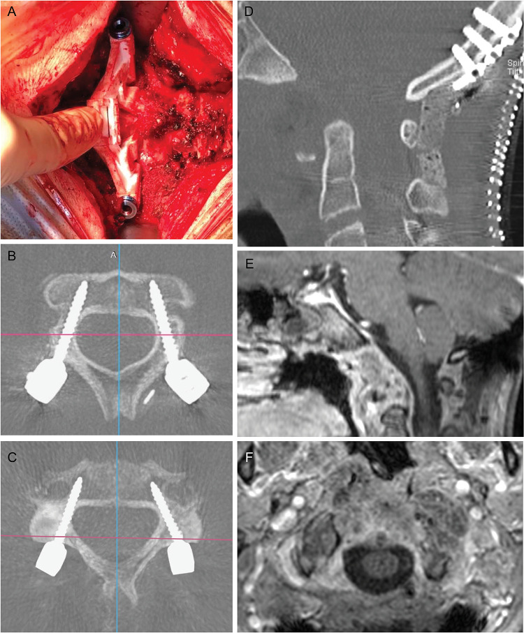
Figure 3.
Intraoperative view of the printed guide fitting on C3 lamina (A) and postoperative results. Transversal CT scan of screws' trajectories in C2 and C3, respectively (B, C). The result of anterior decompression was shown in (D) and an occipital plate and bicortical screws connected with precontoured rods completed the occipitocervical constructs. One month brain and spine MRI (E, F) confirms the decompression of the cervicomedullary junction and the optimal extent of resection.

