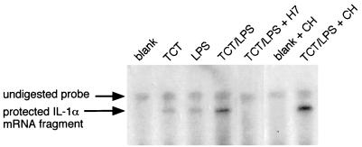
FIG. 2.
TCT-LPS induction of IL-1α mRNA. HTE cells were exposed to TCT (3.2 μM), LPS (100 eU/ml), cycloheximide (CH; 10 μM), and H7 (100 μM) in serum-free medium as indicated. After 4 h, total RNA was prepared and subjected to RPA analysis for IL-1α. (A small remnant of undigested probe remains in all samples due to incomplete degradation of the DNA template used to transcribe the labeled RNA probe.) The protected IL-1α fragment runs slightly lower than the probe, due to noncomplementary vector sequence present at one end of the probe.

